https://iu.pressbooks.pub/humanphys/chapter/cardiac-pacemaker-and-muscle-cells/

https://open.oregonstate.education/aandp/chapter/19-2-cardiac-muscle-and-electrical-activity/
Pacemaker Cells:
These specialized cells are responsible for initiating and regulating the rhythmic contractions of the heart.
Only about one percent of the heart muscle cells are pacemaker cells, while the rest are contractile cells.
Unlike skeletal muscles and neurons, pacemaker cells do not have a stable resting potential. Instead, they exhibit a phenomenon called autorhythmicity.
Here’s how it works:
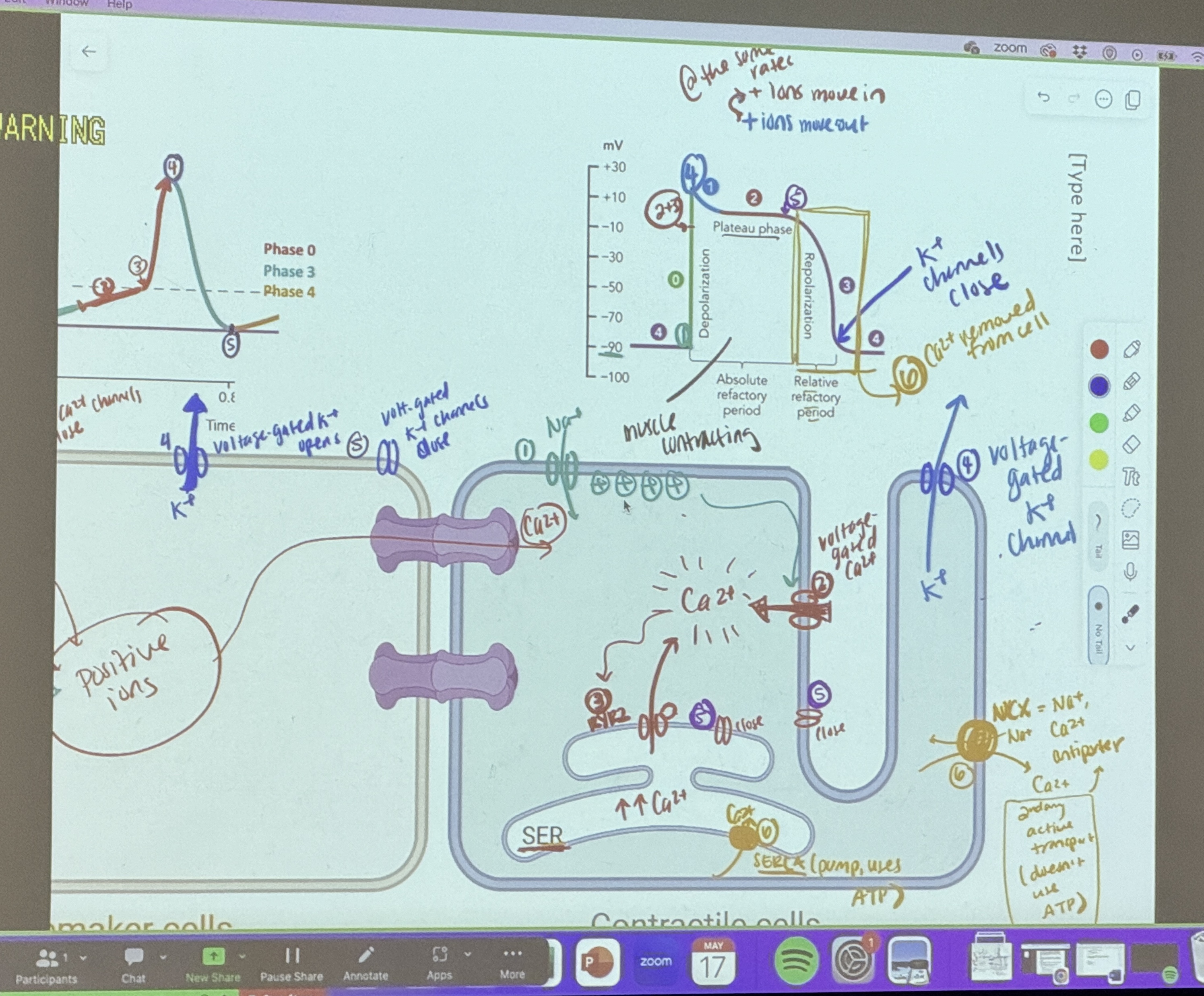
Pacemaker cells contain a series of Na+ channels that allow a slow influx of sodium ions, causing the membrane potential to rise gradually from an initial value of around -60 mV to about -40 mV.
This slow depolarization brings the cell closer to the threshold.
When the threshold is reached, voltage-gated Ca2+ channels open, leading to rapid depolarization until the membrane potential reaches approximately +5 mV.
At this point, the Ca2+ channels close, and voltage-gated K+ channels open, allowing potassium ions (K+) to flow out, leading to repolarization.
Once the membrane potential reaches approximately -60 mV again, the K+ channels close, and the slow Na+ channels reopen, initiating the drift phase once more.
The lack of a stable resting potential in pacemaker cells accounts for their ability to spontaneously depolarize and contract, setting the pace for the heart’s rhythm1.
Contractile Cells:
These cells are responsible for the forceful contractions that propel blood throughout the body.
Their action potential differs significantly from that of pacemaker cells:
Rapid depolarization occurs, followed by brief repolarization and plateau phases.
The plateau phase is crucial for the long refractory periods required for effective blood pumping by cardiac muscle cells before they can fire again.
Contractile cells are connected to neighboring pacemaker cells via gap junctions, allowing them to locally depolarize adjacent cells2.
In summary:
Pacemaker cells initiate and regulate the heart’s rhythmic contractions.
Contractile cells generate forceful contractions to pump blood throughout the body. Together, they orchestrate the intricate dance of the heart’s beating rhythm! 🫀🎵34
Learn more the heart’s beating rhythm! 🫀🎵34
1
iu.pressbooks.pub
2
en.wikipedia.org
3
thisvsthat.io
4
pediaa.com