Wednesday, June 19, 2024
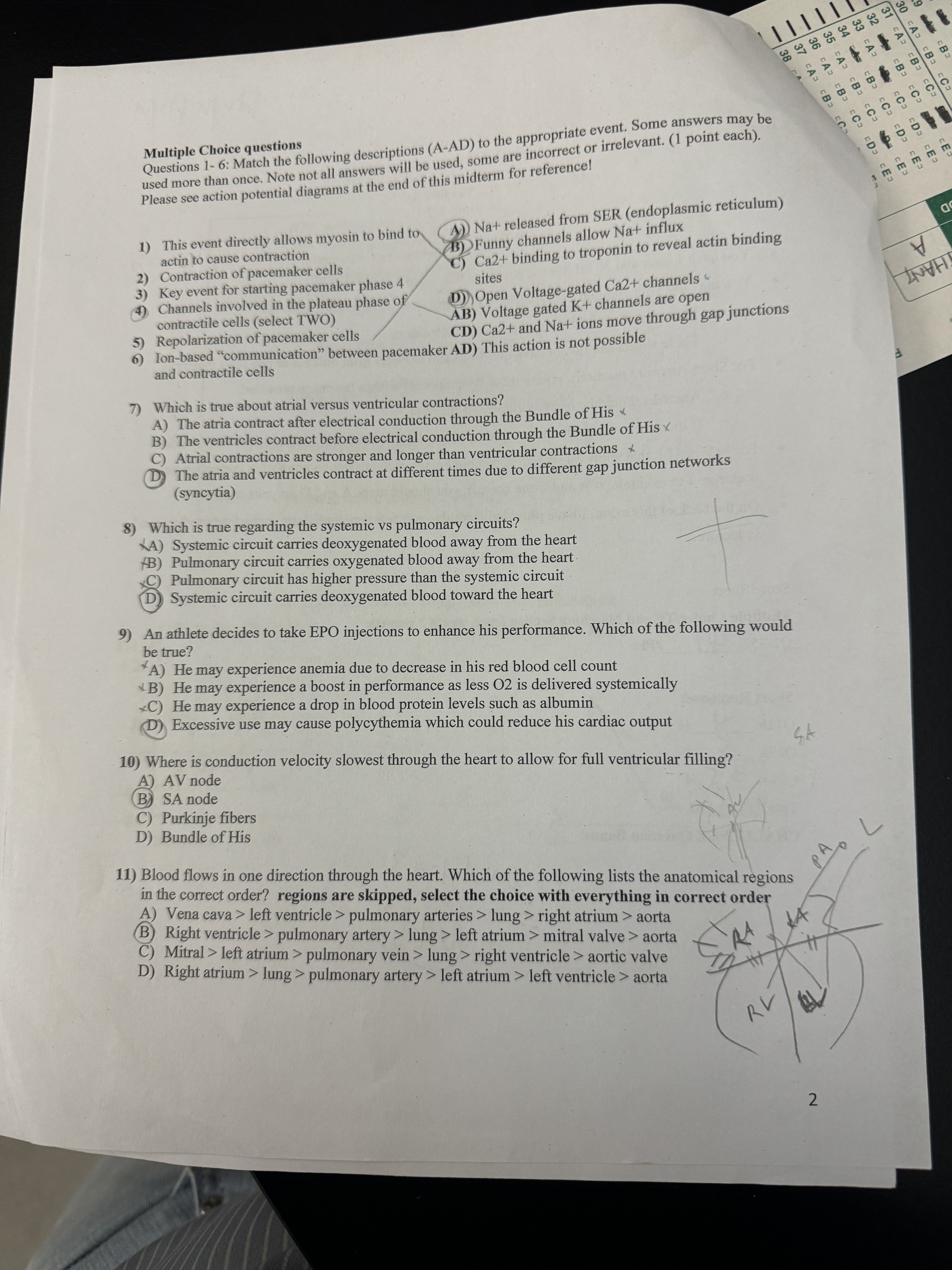
Midterm2

 Chronotropic drugs may change the heart rate and rhythm by affecting the electrical conduction system of the heart and the nerves that influence it, such as by changing the rhythm produced by the sinoatrial node.
 - Positive chronotropes increase heart rate; negative chronotropes decrease heart rate.
- A dromotrope affects atrioventricular node (AV node) conduction.
- A positive dromotrope increases AV nodal conduction, and a negative dromotrope decreases AV nodal conduction.
- A lusitrope is an agent that affects diastolic relaxation.
Many positive inotropes affect preload and afterload.


What does increased CO2 do to blood?
As levels of CO2 in the blood begin to rise, the body can respond through hyperventilation or hypoventilation, respectively.
The CO2 that is bound to hemoglobin forms a carbamino compound. In circumstances where the CO2 and H+ concentrations are high, the affinity of hemoglobin for O2 is decreased.
Second, carbon dioxide can bind to plasma proteins or can enter red blood cells and bind to hemoglobin.
This form transports about 10 percent of the carbon dioxide.
When carbon dioxide binds to hemoglobin, a molecule called carbaminohemoglobin is formed. Binding of carbon dioxide to hemoglobin is reversible.
Hypercapnia occurs when oxygen and CO2 levels become imbalanced in the bloodstream. This imbalance changes the pH balance of your blood, making it too acidic. This condition is called metabolic acidosis. Metabolic acidosis can put excess strain on the kidneys, which can lead to kidney disease or failure. Mar 3, 2023
Carbon dioxide (CO2) has a profound and reversible effect on cerebral blood flow, such that hypercapnia causes marked dilation of cerebral arteries and arterioles and increased blood flow, whereas hypocapnia causes constriction and decreased blood flow [167,168].
The Effects of Second-Hand Tobacco Smoke
EVERYDAY CONNECTION
The Effects of Second-Hand Tobacco Smoke
The burning of a tobacco cigarette creates multiple chemical compounds that are released through mainstream smoke, which is inhaled by the smoker, and through sidestream smoke, which is the smoke that is given off by the burning cigarette. Second-hand smoke, which is a combination of sidestream smoke and the mainstream smoke that is exhaled by the smoker, has been demonstrated by numerous scientific studies to cause disease. At least 40 chemicals in sidestream smoke have been identified that negatively impact human health, leading to the development of cancer or other conditions, such as immune system dysfunction, liver toxicity, cardiac arrhythmias, pulmonary edema, and neurological dysfunction. Furthermore, second-hand smoke has been found to harbor at least 250 compounds that are known to be toxic, carcinogenic, or both. Some major classes of carcinogens in second-hand smoke are polyaromatic hydrocarbons (PAHs), N-nitrosamines, aromatic amines, formaldehyde, and acetaldehyde.
Tobacco and second-hand smoke are considered to be carcinogenic. Exposure to second-hand smoke can cause lung cancer in individuals who are not tobacco users themselves. It is estimated that the risk of developing lung cancer is increased by up to 30 percent in nonsmokers who live with an individual who smokes in the house, as compared to nonsmokers who are not regularly exposed to second-hand smoke. Children are especially affected by second-hand smoke. Children who live with an individual who smokes inside the home have a larger number of lower respiratory infections, which are associated with hospitalizations, and higher risk of sudden infant death syndrome (SIDS). Second-hand smoke in the home has also been linked to a greater number of ear infections in children, as well as worsening symptoms of asthma.
Gas exchange in pulmonary
Once the blood is oxygenated, it drains from the alveoli by way of multiple pulmonary veins, which exit the lungs through the hilum.
alveolar wall consists of three major cell
The alveolar wall consists of three major cell types: type I alveolar cells, type II alveolar cells, and alveolar macrophages.
the Respiratory System
https://www.radiologymasterclass.co.uk/tutorials/chest/chest_home_anatomy/chest_anatomy_page2
(anatomy quiz https://www.kenhub.com/en/library/anatomy/hilum-of-the-lung
two major sections: the external nose, and the nasal cavity or internal nose.
Several bones that help form the walls of the nasal cavity have air-containing spaces called the paranasal sinuses, which serve to warm and humidify incoming air.
Sinuses are lined with a mucosa.
Each paranasal sinus is named for its associated bone: frontal sinus, maxillary sinus, sphenoidal sinus, and ethmoidal sinus.
pharyngeal tonsil, also called an adenoid, is an aggregate of lymphoid reticular tissue similar to a lymph node that lies at the superior portion of the nasopharynx.
The oropharynx is a passageway for both air and food.
The oropharynx is bordered superiorly by the nasopharynx and anteriorly by the oral cavity.
A type II alveolar cell is interspersed among the type I cells and secretes pulmonary surfactant, a substance composed of phospholipids and proteins that reduces the surface tension of the alveoli.