Thursday, May 30, 2024

 
sidebar interaction. Press tab to begin.

In previous quarters where I teach 2 lectures a week in person, the students learn about different capillaries before this lecture. This quarter, this is your pre-class work so it's okay if you didn't know the answer before I covered it!

I'll link a short video on the different types of capillaries for you to review if needed (see it under the instructions for this assignment in "extra resources"

9 regions


Electrocardiography (ECG/EKG) - basics

Arrhythmias - What is an arrhythmia and how is it treated?

The different types of heart arrhythmia

Wednesday, May 29, 2024

two different pressures

physio flip

muscle or nervous tissue 

capillary wit hthe thin wall exchange nutrients

get stuff into the blood 

get stuff out of the blood 

swell up -edema 

fluid stay outside the blood  vessel 

normally reabsorb 

fluid been out of the blood 

hydrostatic pressure 

straw - fill up the straw with water ( water is being pull down by hydrostatic pressure ) 

basic the same thing happen in the capillaries

in the capillaries-CVS system fluid is force out through capillary wall 

just about all the time 

osmotic pressure 

water always flow towards 

higher concentration 

small hole - allow small perticle to pass 

much higher 

protein molecule 

concentration protein molecule 

water always flow 

water reabsorb to the vessel

blood serum protein compre outside the 

driven by osmotic or osmotic pressure ( tend to drive back in ) 

colloid -oncotic pressure 


protein 

ion sodium 

do not contribute the osmotic fluid 

filter 

reabsorb 

osmotic 

interstitial 

capillary 

equal to 

hydrostatic pressure 

small fraction 

final player 

lympatic system 

helping gup prevent or  

Monday, May 27, 2024

 sympathetic nervous system increase Beta 1 adrnergic receptors on contractile cell 

calcium can combine to troponin tropomyosin which mean that we can get stronger contraction or contractile cell increase contraction 

less calcium flow in  ( concentration gradient distrupt ) so that not true 

hyperkalemia tends to block depolarization ( pacemaker cell ) K want to leave the cell 

not contract contractile cell not depolarize

 preload 

contract 

all the step of the cardiac cycle

diastolic volume 

systolic volume


two cardiac out put 

regular stroke volume '

remember your rule 

big picture 

blood flow from high to low 

all about pressure 

cotracting either atria or ventricle 

get to the lung 

get out  from the lung 

valve 

prevent back from 

when ventricle  are ontracting 

prevent from improper blood flow 

all the blood pump out to the body

definition 

  1. stroke volume: the volume of blood pump out the left ventricle of the heart during each systolic cardiac contraction (SV)
  2. end diastolic volume (EDV) the amount of blood that is in the ventricles before the heart contracts 
  3. the volume of blood in one ventricle at the end of the systolic ejection phase immediately before diastole (ESV)
  4. the amount of blood pumped by the heart minute by one ventricle (cardiac output )

5-6 liter of pump 

heart rate 

10 L 

how much blood 

3 key factor 

preload -how much fill the ventricle during diastolic 

force of contract- the more blood push

 - after load -certain amount of resistant 

ventricle has to overcome out through the aorta

second-Heart rate 

Heart rate is higher - the more blood volume to the heart 

if we increase - cardiac output will be increase 

double heart rate 

equation - double the cardiac output 

average heart rate 70 to 72 perminute 

18-40years of age 

infant and toddler

normal average is faster than adult (100-150)

greater than 60 years old 

number of beat per minute 

range from 100 to 130 per minutes 

how heart rates changes 

brady cardia 

slower heart rate 

for average adult ( less than 60perminute ) 

lower than 60 when sleeping heart rate can be slow down 

slower heart rate

the other term 

tachy cardia 

fast than normal heart rate 

over the 100 beat per minute (during exercise to deliver more oxygen ) 

higher metabolic activity

especially aerobic or running 

blood is red 

second set of exercise 

effectors heart rate in two ways - 

positive chronotropic effector- increase the heart rate 

negative chronotropic effector - decrease the heart rate 

tachycardia - decrease preload (fast heart rate ) filled before ventricle contract 

ventricle never fully filled 

start to contract before fully full 


Cardiovascular System Physiology - Cardiac Output (stroke volume, heart ...

Muscle contraction


Sunday, May 26, 2024

CPR can result in broken ribs

 When performed by untrained or overzealous individuals, CPR can result in broken ribs or a broken sternum, and can inflict additional severe damage on the patient. It is also possible, if the hands are placed too low on the sternum, to manually drive the xiphoid process into the liver, a consequence that may prove fatal for the patient. Proper training is essential.

“Staying Alive,” song recorded in 1977 by the Bee Gees

 “Staying Alive,” recorded in 1977 by the Bee Gees. If you are unfamiliar with this song, a version is available on www.youtube.com. 

Subvalvular apparatus: The papillary muscles and the chordae tendineae, known as the subvalvular apparatus, hold the valves closed so that they do not prolapse.

Papillary muscles, together with the chordae tendineae, make up the subvalvular apparatus. 

Blood flows through an atrioventricular (AV) valve Blood flows through an atrioventricular (AV) valve when blood pressure in the atria becomes high during atrial systole and blood pressure in the ventricles becomes low enough during ventricular diastole, creating a blood pressure gradient.

Papillary muscles, finger-like projections from the wall of the ventricles, connect the chordae tendineae (heartstrings) to the cusps of the atrioventricular valves. This connection prevents the valve from prolapsing under pressure.


 Guiding Questions

Cardiac Cycle: Conduction & Mechanical Events


Explain how pressure gradients drive blood flow in the cardiovascular system.

Ventricular contraction ejects blood into the major arteries, resulting in flow from regions of higher pressure to regions of lower pressure.

Identify the location of high pressure and low pressure during systole and diastole.

Outline the steps of the cardiovascular cycle and describe what happens to pressure and blood flow in each chamber at each step of the cycle. Be sure to identify the status of the AV and semilunar valves during each stage of the cycle.

Explain how the pressure gradients cause the valves open and close during the cardiac cycle.

What is the importance of the gap junctions that connect the autorhythmic cells with the contractile cells?

Outline the steps of the intrinsic conducting system and associate them with the steps of the cardiac cycle.

Describe how the SA node controls heart rate. What happens when the SA node is damaged?

Explain how heart rate is controlled by the parasympathetic system and the sympathetic system. Be sure to discuss the intrinsic heart rate, the neurotransmitters and receptors that operate during control of heart rate.

What is an ECG and what kind of information can be gathered from it?

Draw an ECG trace. Identify the P wave, the QRS complex, and the T wave.

Draw another ECG trace. Identify the part of the ECG trace that correlates with atrial depolarization, atrial hyperpolarization, ventricular depolarization and ventricular hyperpolarization.

Now draw a third trace. Identify the part of the ECG trace that correlates with atrial systole, atrial diastole, ventricular diastole, and ventricular systole (remember that physical events happen AFTER the electrical events).

Describe what is happening to both pressure and volume during each stage of the cardiac cycle. Be sure to use the following terms: Ventricular filling, end diastolic volume, isovolumetric contraction, ejection, and end systolic volume, isovolumetric relaxation.

How does ESV change during exercise?

What is stroke volume? What is the average stroke volume? How does stroke volume change?


Cardiac Output


Define cardiac output. How do I determine cardiac output?

Explain how altering heart rate (via sympathetic or parasympathetic stimulation) alters cardiac output.

What is afterload and how does it affect cardiac output? Give an example of how afterload alters cardiac output. 

What is preload and how does it affect cardiac output? Why does EDV determine the force of contraction?

What are some ways that I can alter EDV (and therefore alter stroke volume)?

Describe how the skeletal muscle pump and the respiratory pump affect EDV **(we'll cover this when we discuss blood vessels)

How does total blood volume affect EDV?

How much of the blood is contained in the venous circulation? How can I send more of the blood in the veins to the heart, and how does this affect EDV? **(we'll cover this when we discuss blood vessels)

Define contractility and explain how an inotropic agent affects contractility.

Explain how the sympathetic system modulates contractility. Be sure to name the neurotransmitters and receptors involved.

In a fight or flight response, explain what happens to the following factors and why they are affected this way (be sure to discuss neurotransmitters and receptors): heart rate, venous return, EDV, contractility, stroke volume, cardiac output.

Saturday, May 25, 2024

The Cerebellar GABAAR System as a Potential Target for Treating Alcohol Use Disorder

 The Cerebellar GABAAR System as a Potential Target for Treating Alcohol Use Disorder 

The Cerebellar GABAAR System as a Potential Target for Treating Alcohol Use Disorder

refractory period


 The refractory period is a state of recovery that occurs after a neuron has fired an action potential. 
During this period, another action potential cannot be easily produced. 
This encourages unidirectional flow of action potentials because they cannot travel backwards to inactive neurons.

The depolarization that produces Na+ channel opening also causes delayed activation of K+ channels and Na+ channel inactivation, leading to repolarization of the membrane potential as the action potential sweeps along the length of an axon (see Figure 3.12). In its wake, the action potential leaves the Na+ channels inactivated and K+ channels activated for a brief time. These transitory changes make it harder for the axon to produce subsequent action potentials during this interval, which is called the refractory period. Thus, the refractory period limits the number of action potentials that a given nerve cell can produce per unit time. As might be expected, different types of neurons have different maximum rates of action potential firing due to different types and densities of ion channels. The refractoriness of the membrane in the wake of the action potential explains why action potentials do not propagate back toward the point of their initiation as they travel along an axon.

What happens during the refractory period of a neuron?
In its wake, the action potential leaves the Na+ channels inactivated and K+ channels activated for a brief time. These transitory changes make it harder for the axon to produce subsequent action potentials during this interval, which is called the refractory period.
By definition, the refractory period is a period of time during which a cell is incapable of repeating an action potential. In terms of action potentials, it refers to the amount of time it takes for an excitable membrane to be ready to respond to a second stimulus once it returns to a resting state.

Friday, May 24, 2024

Aorta

 The aorta holds the distinction of being the largest artery in the human body. Its significance lies in serving as the initial pathway for blood leaving the heart and distributing it to various organs through smaller, branching arteries. Without the aorta, our body’s tissues wouldn’t receive the vital oxygen and nutrients they need 1. Let’s delve into more details about this remarkable vessel:

  • Anatomy of the Aorta:

    • The aorta starts at the left ventricle of the heart, where it receives oxygenated blood.
    • It then arches upward toward the neck (forming the aortic arch), before curving back downward and extending into the abdomen.
    • Structurally, the aorta is a big tube-like structure, typically about 1 inch wide in diameter. However, its size can vary based on an individual’s height and weight.
    • The aorta is widest at its connection to the heart’s left ventricle through the aortic valve. As it descends into the abdomen, it gradually narrows.
  • Four Parts of the Aorta:

    1. Ascending Aorta:
      • The first part of the aorta, connected to the left ventricle.
      • Originates at the aortic valve, which regulates blood flow from the heart into the aorta.
    2. Aortic Arch:
      • Curves upward toward the head and neck.
      • Begins where the ascending aorta ends.
    3. Descending Aorta:
      • Travels downward from the aortic arch through the chest.
      • Also known as the descending thoracic aorta.
    4. Abdominal Aorta:
      • The final part of the aorta.
      • Starts at the diaphragm and ends where it splits into the two iliac arteries that extend into the legs.
  • Layers of the Aorta Wall:

    • The aorta has three layers in its wall:
      1. Tunica Intima: The innermost layer, providing a smooth surface for blood flow. Composed of endothelial cells and supporting tissue.
      2. Tunica Media: The middle layer, consisting of smooth muscle, elastic tissue, and collagen.
      3. Tunica Adventitia: The outer layer made of connective tissue (collagen) and a network of small blood vessels (vasa vasorum) that nourish the aorta.
  • Variations and Anomalies:

    • In some conditions (such as dextrocardia or situs invertus), the aorta may be on the right side instead of the left.
    • Coarctation of the aorta is a birth defect where a part of the aorta is significantly narrower than usual.

In summary, the aorta’s essential role as the largest artery ensures efficient oxygen and nutrient distribution throughout our body, making it a vital conduit for life-sustaining blood flow 234. If you have more questions or need further information, feel free to ask! 😊

lab exit


What Is Aortic Stenosis? Your aortic valve keeps blood flowing from your heart's lower left chamber (left ventricle) to the aorta which is the main artery bringing blood from the heart to the body. Aortic stenosis occurs when the aortic valve narrows and blood cannot flow normally.

regurgitation?
Mitral valve regurgitation is a problem that occurs when there is a valve leak, leading to blood flowing back into the left atrium. The incomplete valve closure may produce a softer S1 sound. Symptoms that may arise in people with this valve problem include: dizziness.


check in question week 7


tetrodotoxin TXX blocks voltage gated sodium channel which part of action potential graph is affected by TXX discuss the activation and inactivation status of channel the key channel involved and the refractory period

 Tetrodotoxin (TTX) is a potent neurotoxin that specifically targets voltage-gated sodium channels. Let’s explore how TTX affects the different phases of the action potential graph:


Activation and Inactivation of Sodium Channels:

Voltage-gated sodium channels play a crucial role in generating action potentials.

These channels have two main states: closed (resting state) and open (activated state).

When a neuron is at rest, sodium channels are predominantly in the closed state.

Upon depolarization (due to an incoming stimulus), some sodium channels open, allowing sodium ions (Na⁺) to flow into the cell. This is the activation phase.

However, sodium channels also have an inactivation gate that can close the channel shortly after it opens. This prevents continuous sodium influx.

Tetrodotoxin’s Mechanism of Action:

TTX binds to the extracellular pore opening of voltage-gated sodium channels.

By binding to site 1 of the sodium channel, TTX temporarily disables the channel’s function.

Specifically, it prevents the passage of sodium ions into the neuron during the rising phase of an action potential.

As a result, the nervous system cannot transmit messages effectively, leading to muscle paralysis.

Key Channel Involved:

The primary sodium channel affected by TTX is the fast voltage-gated sodium channel.

These channels are responsible for the rapid depolarization phase of the action potential.

Refractory Period:

The refractory period is a critical aspect of action potentials.

After an action potential, sodium channels enter a refractory state:

Absolute refractory period: During this phase, sodium channels are completely closed and cannot be activated, regardless of the stimulus.

Relative refractory period: In this phase, some sodium channels have recovered from inactivation, but the membrane potential is still below the resting level. A stronger stimulus can trigger another action potential.

TTX prolongs the refractory period by preventing sodium channels from reopening promptly.

In summary, TTX disrupts the normal function of sodium channels, leading to the inhibition of action potentials and muscle paralysis. Its selective blocking action makes it a valuable tool for studying neuronal excitability and pain pathways12. 🧪🔬


If you have more questions or need further clarification, feel free to ask! 😊

what is the concentration gradient for the ion at the resting membrane potential what happen ion flow once the drug added and how the drug change the memembrane potential

 what is the concentration gradient for the ion at the resting membrane potential what happen ion flow once the drug added and how the drug change the memembrane potential


Copilot

Certainly! Let’s break down the impact of a drug on the resting membrane potential (RMP) and how it affects ion flow.


Concentration Gradient at RMP:

At the resting membrane potential, there is a concentration gradient for both sodium ions (Na⁺) and potassium ions (K⁺).

Inside the cell, the concentration of potassium ions is higher than outside, while the concentration of sodium ions is higher outside the cell.

This concentration gradient is maintained by the sodium-potassium pump and leak channels.

Ion Flow Once the Drug Is Added:

When a drug is introduced, it can alter the permeability of the cell membrane to specific ions.

Let’s consider a hypothetical drug that selectively increases the permeability of sodium ions (Na⁺) through the membrane.

Impact of the Drug on Membrane Potential:

If the drug enhances sodium ion permeability, more sodium ions will flow into the cell.

This influx of positive charges will depolarize the cell membrane (make it less negative).

As a result, the resting membrane potential will shift toward a less negative value (e.g., from -70 mV to a less negative value).

Overall Effect:

The drug disrupts the balance between sodium and potassium ions, altering the resting membrane potential.

Depending on the drug’s mechanism, it could lead to hyperpolarization (more negative potential) or depolarization (less negative potential).

Remember that the specific effects of a drug depend on its properties and the type of ion channels it interacts with. Real-world drugs can have complex interactions, affecting various ion channels and receptors.


If you have any further questions or need additional details, feel free to ask! 🧪🔬

explain how each of the choices selected above RMP K leack channel and sodium and potassium pump and concentration gradient and membrane permeablity why RMP is -70


the factors that contribute to the resting membrane potential (RMP) of a neuron and why it is typically around -70 mV.


Leak Channels:

Neurons have leak channels (also known as non-gated channels) that allow ions (such as potassium and sodium) to passively move across the cell membrane.

These channels are always open and contribute to the resting membrane potential.

The most important leak channel for RMP is the potassium leak channel (K⁺ channel). It allows potassium ions (K⁺) to move out of the cell, making the inside of the cell more negative.

Sodium-Potassium Pump (Na⁺/K⁺ Pump):

The sodium-potassium pump actively transports sodium ions (Na⁺) out of the cell and potassium ions (K⁺) into the cell.

For every three sodium ions pumped out, two potassium ions are pumped in.

This pump helps maintain the concentration gradients of sodium and potassium across the cell membrane, which is crucial for RMP.

Concentration Gradient:

The concentration gradient refers to the difference in ion concentrations between the inside and outside of the cell.

At rest, there is a higher concentration of potassium ions (K⁺) inside the cell and a higher concentration of sodium ions (Na⁺) outside the cell.

The potassium leak channel allows potassium ions to move out of the cell along their concentration gradient, contributing to the negative RMP.

Membrane Permeability:

The permeability of the cell membrane to different ions affects RMP.

The cell membrane is more permeable to potassium ions (K⁺) due to the abundance of potassium leak channels.

Sodium ions (Na⁺) have fewer leak channels, so their contribution to RMP is smaller.

Now, let’s tie it all together:


The potassium leak channel allows potassium ions to leak out of the cell, making the inside more negative.

The sodium-potassium pump maintains the concentration gradients by actively pumping sodium out and potassium in.

The net effect of these processes results in an RMP of approximately -70 mV.

Remember that RMP can vary slightly depending on the specific type of neuron and its environment, but -70 mV is a common value. 


If you have any more questions or need further clarification, feel free to ask! 😊

Thursday, May 23, 2024

 The first heart sound "lubb" (or S1) corresponds to AV valves closing. The second heart sound "dubb" (or S2) corresponds to the closing of semilunar valves closing.


The closing of these valves are so forceful that you can hear these when you are listening to heart beats of a person/patient and this is what we refer to when we are listening to someones heart beat!

Answer 1:
Answer 2:

The Cardiac Cycle, Animation

Cardiac Cycle | Wiggers Diagram | Part 5 | Summary | Cardiac Physiology

Muscle Contraction - Cross Bridge Cycle, Animation.

Lecture 6: Cardiac Cycle Conduction Friday 5/17

 Lecture 6: Cardiac Cycle Conduction

Friday 5/17


Pre-Class SlidesDownload Pre-Class Slides

Take notes with me using this handoutDownload Take notes with me using this handout

Lecture 6 (5/17) Exit Ticket (opens when I open it during class!)

Post-class slidesDownload Post-class slides

RecordingLinks to an external site.

This is actually a really nice summary of the whole cardiovascular section we'll cover over the next few weeks. For pre-nursing folks, this is actually put together for their Associate degree in nursing (so this stuff is quite relevant!). I'll leave this here in case folks like a one-stop resource of helpful diagrams & text. Otherwise feel-free to review lecture recordings & pre-class videos: https://www.austincc.edu/apreview/PhysText/Cardiac.htmlLinks to an external site.

Lecture 10 handout Key (will post at the end of the week :) have some folks making up the lecture)


Guiding Questions:


1. Lay out the steps of myocardial cell contraction. Describe carefully what happens in each step.


What is the Ca2+ spark (rush of calcium into the cell)? Where does the Ca2+ come from?

Lay out the steps that lead to relaxation of the myocardial cell after contraction.

How is Ca2+ removed in a contractile myocardial cell?

3. What is the resting membrane potential of a contractile myocardial cell?


What is the trigger for the action potential in contractile myocardial cells? What is the source of that stimulus?

What type of channel activity results in the rising phase, plateau phase and falling phase of the action potential? What is the trigger for each of these types of channel activity?

4. Why is the refractory period in contractile cells so much longer than that in skeletal muscle cells? (covered in post-class week 6, not lecture)


How does the refractory period for myocardial cells prevent summation of force in these cells*****

What would the impact of summation be if it could happen in these cells? Where does the Ca2+ come from to lengthen the refractory period so much? ****These questions will actually be answered during your pre-class week 7! Feel free to skip for now and make sure they are answered by next week!

5. Describe the origin of the pacemaker potential found in autorhythmic cells.


How does the pacemaker potential trigger the rising phase in autorhythmic cells?

What causes the falling phase in autorhythmic cells?

Compare and contrast the rising phase of the autorhythmic cells with the rising phase of the contractile cells, the rising phase in skeletal muscles and the rising phase in neurons.

6. How do action potentials in autorhythmic cells translate to the contraction of the contractile cells?


. What is the function of the cardiovascular system? Give some examples of materials transported by the system.


Describe the organization, pattern of blood flow and the major function of the cardiovascular system using the following terms: heart, arteries, capillaries, and veins.

Describe the structure of the heart and pattern of blood flow through the heart using the following terms: atria, ventricles, septum, vena cava, aorta, pulmonary veins, pulmonary arteries, AV valves, papillary muscle*, chordae tendineae*, semilunar valves.

What is the function of the valves in the heart? Where does the energy come from to open and close the valves? (*covered in lab Week 6)

2. What is the importance of the gap junctions that connect the autorhythmic cells with the contractile cells?


Outline the steps of the intrinsic conducting system and associate them with the steps of the cardiac cycle.

Describe how the SA node controls heart rate. What would happen when the SA node is damaged?

Explain how heart rate is controlled by the parasympathetic system and the sympathetic system. Be sure to discuss the intrinsic heart rate, the neurotransmitters and receptors that operate during control of heart rate

3. Define the terms systole and diastole in terms of the cardiovascular system.****


Explain how pressure gradients drive blood flow in the cardiovascular system. Identify the location of high pressure and low pressure during systole and diastole.*** these questions will be covered next Tuesday's lecture

week 2 preclass activity quizz


Question 2
0 / 1 pts


When I teach this in person, I like to make an analogy between pacemaker cells, contractile cells, and the autonomic nervous system (ANS). Remember that the ANS is divided into sympathetic (speeds up heart rate) and parasympathetic (slows down heart rate).


Consider how lights can be turned on/off and that some have a dimmer switch:

dimmer.webplights .webp


I would argue that each component: pacemaker, contractile cells and autonomic nervous system match the way this lighting system works. Check your understanding by matching the terms below.

Correct!
Pacemakers are autorhythmic and determine when contractile cells will depolarize.
 
Correct!
ANS can determine how fast or slow pacemaker and contractile cells depolarize and therefore speed up/slow down heart rate.
 
Correct!
Contractile cells are THE cardiac muscle cells that generate contractions (pumping/beating) of the heart. They follow the pace set by pacemakers & ANS.


How are contractile and pacemaker cells connected? Select all

Correct!
  
  
You Answered
  
Correct Answer
  

How does the heart pump? How does it know when to beat?

  
Correct!
  

Wednesday, May 22, 2024

Week 3 practice question

 Question 1

1 / 1 pts

Chlorpromazine (marketed under brand names Thorazine and Largactil) is a drug used to treat psychotic disorders such as schizophrenia or manic-depression in adults. Chlorpromazine was developed in the 1950s and its introduction as the first antipsychotic on the market has been labeled as one of the great advances in the history of psychiatry.

Chlorpromazine blocks postsynaptic receptors for several different neurotransmitters, such as dopamine and serotonin. Predict the effect of chlorpromazine on synaptic transmission by selecting all of the steps it will directly inhibit in the picture below.

Screen Shot 2022-08-26 at 10.45.58 AM.png


Question 2
1 / 1 pts
Some neurotransmitter receptors are ion channels that open when the neurotransmitter binds to them. Those receptors are called:


Question 3
1 / 1 pts

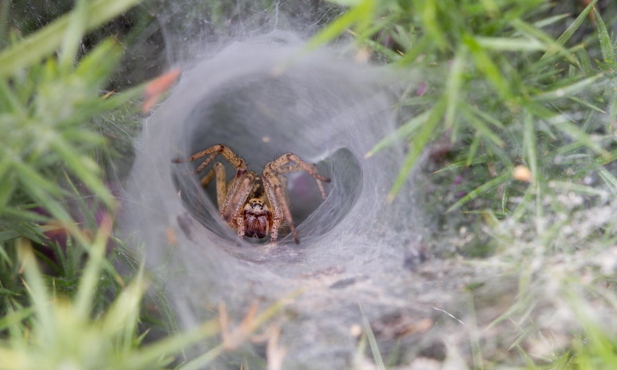
Funnel-web spiders are found in southeast Australia, around Sydney. Others are found in Europe, New Zealand, and Chile. They are not native to the United States, although some people may keep them as exotic pets.
Their bites are very painful and dangerous. They have been known to cause serious symptoms, such as agitation, confusion, numbness of mouth and lips, and even coma and death.

Funnel-web spider venom is a neurotoxin that blocks gated calcium channels. Its effect would be a/an:


Question 4
1 / 1 pts

Identify the ion that would move through the channel to produce an IPSP.

Ions 1-3 are in the extracellular fluid (blue background),  ions 4-6 are in the cytosol (yellow background). Figure out which ion and which direction it would flow based on what you know about concentration gradients for each ion.

Screen Shot 2022-08-26 at 10.53.19 AM.png


Question 5
1 / 1 pts
One type of channel involved in generating postsynaptic potentials at a chemical synapse is the:


Question 6
1 / 1 pts
Screen Shot 2022-08-26 at 11.01.39 AM.pngSelective serotonin reuptake inhibitors (or SSRIs), such as Prozac, Celexa, Lexapro, Paxil, and Zoloft, are a class of drugs used to treat mood disorders such as depression and anxiety. They inhibit the transporter responsible for serotonin reuptake into the presynaptic neuron. What is the effect of the SSRIs on the serotonin concentration in the synaptic cleft?
  
Correct. When reuptake is inhibited, more serotonin stays in the cleft longer and is available to stimulate postsynaptic neurons. Since mood disorders often correlate with reduced serotonin availability and receptor stimulation, SSRIs are effective in helping patients who suffer from these disorders.
  
 
Question 7
1 / 1 pts
Which of the following does NOT happen to neurotransmitters during synaptic transmission termination?


Question 8
1 / 1 pts

Use what you learned during the Lecture 3 handout.

Cocaine is a powerfully addictive stimulant drug made from the leaves of the coca plant native to South America. As a street drug, cocaine looks like a fine, white, crystal powder or it is processed to a make rock crystal.

Cocaine acts as a dopamine reuptake inhibitor. Therefore, cocaine will:


Correct. Less reuptake means more dopamine remains in the cleft and is available to stimulate dopamine receptors.


Question 9
1 / 1 pts
During the absolute refractory period, the axon membrane:


Question 10
1 / 1 pts
The refractory period of an action potential...


Question 11
1 / 1 pts
The faster, more efficient way of propagating an action potential is:


Question 12
1 / 1 pts
The threshold potential is best defined as the potential at which...


Question 13
1 / 1 pts
Action potentials self-propagate because...